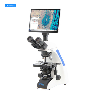
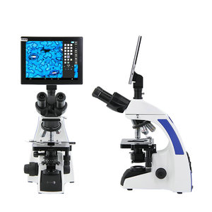
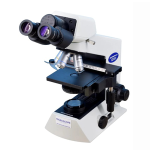
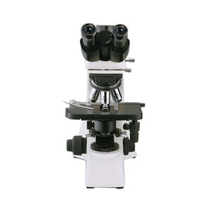
Types of biological microscopes with camera
The market provides users with many options for a biological microscope with a camera. Each of these types is destined for a specific purpose. Therefore, it is very important that one chooses wisely. The following list goes over the most common biological microscopes with cameras.
Flash Lamp Biological Microscope with Camera
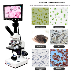
This type of biological microscope uses the illumination power of a flash lamp. The flash lamp provides illumination, and then the camera attached to the microscope takes images. This apparatus is suitable for observing various biological specimens that require a microscope for precise observation. These specimens can range from cells, tissues, microorganisms, plant samples, etc. Another good thing about such a microscope is that it is portable. This factor helps people to conduct field research without any power source. The compact design also makes the storage of this instrument simple and easy.
Live Cell Biological Microscope with Camera
This camera-equipped biological microscope is mainly used in biological research. This is because it allows scientists to observe live cells in real time. Doing so enables researchers to monitor cellular processes, movements, and interactions vital for cellular biology. To enable the viewing of live samples, this type of biological microscope has to bring into focus effective and advanced imaging tactics like phase contrast, fluorescence, or confocal microscopy.
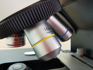
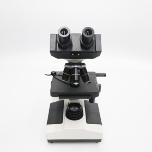
High Power Lens Biological Microscope with Camera
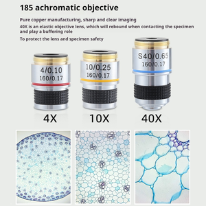
A high-power lens biological microscope uses high magnification objective lenses to view minute specimens. It is especially useful for detailed observations of biological samples like tissues and cells. This equipment particularly suits those engaged in detailed histology or cytology studies.
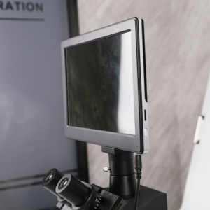
LED Lamp Biological Microscope with Camera
An LED biological microscope with a camera merges optics and electronics. The illumination is done using an LED, while the image capture is done by a camera. The camera allows the users to view the specimen in real time. Also, it enables the user to take high-quality photographs. The microscope has a long working life due to the energy-efficient nature of LED. This makes it suitable for long observation sessions, too, as it does not overheat.
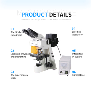
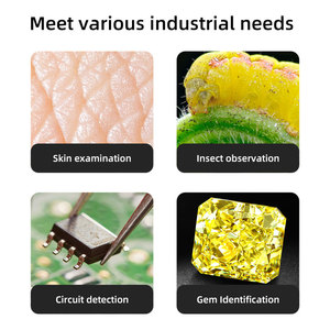
Industrial applications of biological microscopes with camera
Biological microscopes with cameras have become a huge necessity in various operations. Such operations span scientific research, healthcare, education, and even environmental studies. Here is a rundown of some industrial applications of these devices.
Microbiology
In this field, cameras on biological microscopes allow for the detailed imaging of bacteria, fungi, and other microorganisms. These microorganisms are then recorded for research and educational purposes. The detailed imaging capabilities of cameras also contribute to more accurate and informative microscopic studies.
Cell Biology
To study cell structures and functions, cell biologists strongly rely on biological microscopes with cameras. The tools that come with advanced imaging methods permit live cell imaging and fluorescence microscopy. These allow researchers to record cellular activities in real time. These recorded cellular activities can further be analyzed in cellular mechanics and disease progression studies.
Histology
Tissue examination in histology primarily uses a biological microscope with a camera. This helps cut and stain biological tissue samples to examine them under a microscope. Pathologists then use the captured images for disease diagnosis, research, and academic training. Generally, these images are shared by the camera-equipped biological microscopes with digital technology for telepathology.
Pharmaceutical Industry
Here, quality control and drug development processes use biological microscopes with cameras. This hardware allows for the examination of drug formulations and biological samples. Furthermore, the detailed documentation offered by these cameras is vital for regulatory compliance and reporting.
Environmental Science
Mused in ecology studies and environmental monitoring, cameras on biological microscopes help capture detailed images of biological specimens. Specimens such as soil samples, water microorganisms, and plant life. These images are used for habitat assessment, biodiversity studies, and pollution impact evaluations.
Forensic Science
In forensic laboratories, biological microscopes with cameras analyze biological evidence. This evidence can be blood, hair, or other biological material left at a crime scene. Detailed microscopic images of this material help forensic scientists match DNA, identify suspects, and provide crucial visual evidence in criminal investigations.
Product specifications and features of biological microscopes with camera
Biological microscopes with cameras have some mandatory features for any operation. Spelling out these features along with the product specifications sections helps the reader understand these essential components better. They also provide a better user experience.
Technical specifications
-
Resolution
The resolution of a biological microscope with a camera will depend on the sensor quality of the camera. In this case, the higher the megapixels, the clearer the captured images will be.
-
Optical System
Most biological microscopes with cameras will have an optical system that effectively combines objectives and eyepieces. This is to provide a wider field about the functional image. The optical system here focuses on providing clearer magnified images.
-
Illumination
LED and halogen lamps are the most commonly used illumination sources. Both of these sources have advantages. For instance, LED provides long life while being energy efficient. On the other hand, halogen gives bright and high-contrast images.
-
Digital Connectivity
Most biological microscopes support USB connectivity. This allows easy image uploading. Wi-Fi is also supported on some models to enable instant sharing and remote microscopy function capabilities.
-
Software Compatibility
The software most frequently used is measurement, annotation, and image processing software. This software mainly runs on Windows and Mac operating systems.
-
Power Supply
Microscopes that use LED for illumination mainly derive their power supply from batteries or USB. The same goes for rechargeable LED microscopes: their power can be supplemented by USB.
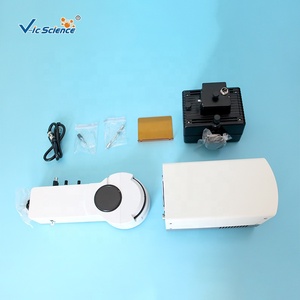
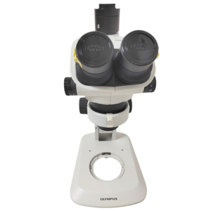
How to install
-
Find a workspace
One should do this because stability is required when using one of these types of microscopes.
-
Mount the base
The first thing one has to do is mount the base of the microscope. The base itself has been designed to provide stability.
-
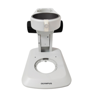
Attach the arm
The arm holds all the important components like the stage and eyepiece. This is why it has to be attached to the base for support.
-
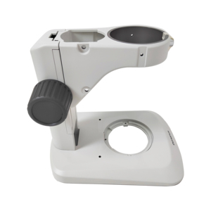
Install the stage
To install the stage, use the provided screws and mounts on the arm to attach it. After that, place the slide holder on the stage.
-
Set the objectives
The lens with the lowest power should be initially positioned near the opening. After doing this, secure the objective lenses into the nosepiece. It is done by rotating the components.
-
Mount the condenser
Attach the condenser to the arm beneath the stage. It should be adjusted to provide better illumination while viewing the slide.
-
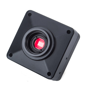
Install the camera
Install the camera on the eyepiece tube. This is because the camera is the main feature of this type of microscope. After attaching the camera, ensure it is secure and aligned properly.
-
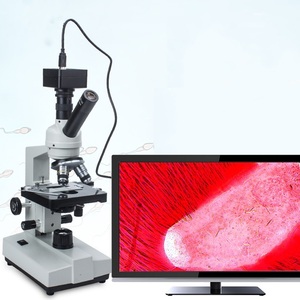
Connect to power and the computer
If the microscope has built-in illumination, connect it to a power source. Connect the camera to a computer via USB. This enables real-time viewing and image capturing.
How to use
-
Prepare the specimen
This is done by placing the biological sample on a glass slide. A coverslip should be placed over the sample to flatten it for better viewing.
-
Place the slide on the stage
The stage of the microscope has slide holders for easy placement of the slides. Just drop the slide in the holders, and it will be in place.
-
Select the objective lens
Spin the nosepiece to the lowest power objective lens. This will help in bringing the sample into a rough view. After doing that, one can switch to a higher power lens for detailed viewing.
-
Adjust the stage
The stage should be lowered using the stage controls to avoid damaging the lens or slide. After it goes low, close the eyepiece. Move the stage up slowly until it comes closer to the slide and locks into place.
-
Illumination
Turn on the microscope's light source after placing the slide on the stage. Adjust the light intensity so it is comfortable to see the specimen and does not cause strain.
-
Focusing
While looking into the eyepiece, use the coarse focus knob to bring the stage into view. After getting the specimen in view, use the fine focus knob to get a clearer and sharper image.
-
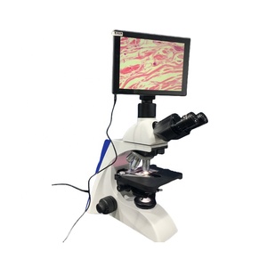
Capturing Images
The last step is to conduct the research. The camera-equipped biological microscope can take high-quality pictures for documentation or study.
Maintenace and repair
-
Regular Cleaning
This is an integral maintenance process. One has to use lens paper to clean the objectives and eyepiece. It should not have any dust or fingerprint for good functioning. The stage and slide holders should be wiped using a damp cloth.
-
Use Coverslips and Proper Mounting
To reduce the contact of specimen with lenses, always use coverslips. Doing this will not only keep the lenses clear but also keep them safe from scratches as well.
-
Avoid Excessive Force
This will ensure microscope parts do not get damaged when in use. Since biological microscopes have different objectives that users must rotate, excessive force may damage these objectives.
-
Regular Inspections
People should always take some time to inspect the hardware. For example, checking focus knobs to ascertain that they work properly.
-
Calibrate Microscope
One way of maintaining a biological microscope's accuracy is by calibrating it regularly. The period might be once every month or according to the frequency of use.
-
Proper Storage
The major storage location for these instruments is dry and dust-free places. In that space, cover the microscope with a dust cover to protect it from dust and debris.
-
Use a Surge Protector
This protects the LED lamp from power surges. Such power surges can cause damage to the internal components of the microscope.
-
Software and Camera Maintenance
The biological microscope with a camera needs regular software updates for better image capture and management. Furthermore, periodic checks on the camera attachment are needed so that it works optimally and smoothly.
Quality and safety factors
Many factors affect the safety of biological microscopes with cameras in industrial applications. On the flip side, they also affect the quality of end products. Here are some of those factors.
Camera Specifications
The quality of the biological images primarily depends on the camera resolution. A camera with high-resolution will capture clear and detailed images. This leads to quality biological research. Microscopes that come with cameras also have internal cameras with different resolutions. Usually, this resolution is measured in megapixels. The higher this number, the more quality the image will have.
Megapixels
Megapixels is the measure of digital images. As stated before, a biological microscope with a camera that has high megapixels will mainly provide clear and sharp images. This will truly help users who need to view minute details of their biological samples. It is particularly important in microbiology and cell biology.
Sensor Size
This is among the most vital factors for the quality of a biological image. A larger sensor size usually captures more light. This is especially advantageous in low-light conditions like fluorescence or live-cell imaging.
Optical Zoom
Optical zoom enhances image quality more than digital zoom. This is because optical zoom uses physical lens elements to magnify a scene. Digital zoom, on the other hand, uses cropping techniques to magnify a scene. Such a process lowers an image's resolution and quality. Go for a biological microscope with a camera that has a high optical zoom.
Data Security
Security issues like unauthorized access to sensitive biological data can be risky. To counter this, digital biological cameras should have strong security measures. These measures include data encryption and secure user authentication. Implementing these measures not only protects confidential data but also complies with regulations like GDPR and HIPAA.
Regular Software Updates
Software is one of the most important parts of life. It too needs updates for the biological microscope to ensure quality and safety. In this world of cyber crime, critical vulnerabilities in software can lead to risks like data breaches. These breaches can cause a lot of financial loss, breaches of trust between the affected party and the victims, and even legal problems.
Quality of Lenses and Filters
Quality lenses and filters ensure accurate and precise imaging of biological samples. This is because they reduce aberrations. Using poor quality will distort the image, so users will end up wasting time due to inaccuracy. High-quality lenses improve the quality of images, whereas high-quality filters improve the quality of live images. This is because it controls the amount of light that passes through the specimen.
Regular Calibration
Regular calibration helps ensure the accuracy of measurements taken during biological experiments. Without it, there will be a decline in measurement accuracy. This decline can have serious repercussions in life sciences, like incorrect drug formulations or misdiagnosed diseases. Users should frequently calibrate their microscopes to maintain its endemic accuracy. Calibration also helps maintain safety standards by ensuring the equipment functions within regulatory parameters.
Q&A
Q1. How does one choose an ideal biological microscope with a camera?
A1.The biological microscope with camera must have good resolution first and foremost. The camera's resolution should be in megapixels. The higher, the better. Its optical zoom power and sensor size also assist in choosing a camera that captures quality images. Afterwards, consider the software compatibility of the camera-equipped biological microscope. This ensures easy image management and analysis. The digital biological microscope must be durable, excellent-performing, and reliable.
Q2. What parts of a biological microscope with a camera should be frequently checked?
A2.Pathological and forensic biological microscopes with cameras should be serviced regularly. During servicing, vital parts like focus knobs, objective lenses, and condensers should be inspected. Other important parts that require inspection and servicing are stage mechanisms and light sources. Inspecting hardware parts boosts the efficiency of these types of biological microscopes. One should also conduct repairs whenever there is damage to any part.
Q3. What impact does software have on the function of biological microscopes with cameras?
A3.Safety and quality software updates ensure the biomedical stage microscope and its camera function securely and effectively. The software manages data captured during experiments for storage and analysis. Users need to update it frequently so the vulnerabilities found in it will be patched.
Q4. What are the common features of a digital biological camera microscope?
A4.This type of biological equipment has USB and Wi-Fi connectivity options as common features. The medical and forensic microscope with cameras also has measurement and analysis software. These features make them a modern gadget for digital microscopy.
Q5. What are some tips for improving the longevity of biological microscopes with cameras?
A5.Status and quality control go a long way in increasing the usefulness of these devices. Also, ensuring that the cameras are handled with care during placements and removals contributes to quality and safety. People should also store this equipment in dry, dust-free environments for enhanced longevity.


 浙公网安备 33010002000092号
 浙B2-20120091-4